Let's Get Started
Let's explore some of the equipment commonly used in laboratory and field investigations in biology. Before you get started, don’t forget to print out your OnTRACK Biology Journal.
TEKS Standards and Student Expectations
B(2) The student uses scientific methods and equipment during laboratory and field investigations. The student is expected to:
B(2)(F) collect and organize qualitative and quantitative data and make measurements with accuracy and precision using tools such as calculators, spreadsheet software, data-collecting probes, computers, standard laboratory glassware, microscopes, various prepared slides, stereoscopes, metric rulers, electronic balances, gel electrophoresis apparatuses, micropipettors, hand lenses, Celsius thermometers, hot plates, lab notebooks or journals, timing devices, cameras, Petri dishes, lab incubators, dissection equipment, meter sticks, and models, diagrams, or samples of biological specimens or structures
Learning Objectives
Identify laboratory and field equipment commonly used by biologists.
Describe when and how laboratory and field equipment are used.
Associate a given measurement with the equipment used to make that measurement.
Essential Questions
What equipment is commonly used in laboratory and field investigations in biology?
What types of measurements do biologists make during investigations and what type of equipment do they use to make these measurements?
Vocabulary
- Ruler
- Vernier Caliper
- Balance
- Graduated Cylinder
- Pipette
- Micropipette
- Dissecting Kit
- Dissecting Microscope
- Compound Microscope
- Hand Lens
- Binoculars
- Gel Electrophoresis
Equipment for Biology: Case Study: Blue Mussel
|
To take a close look at equipment commonly used in biology investigations, let's imagine we are studying the growth, development, and overall health of blue mussels. Blue mussels are shellfish found along shorelines in coastal waters. Biologists often study blue mussels because they are sensitive to marine pollution (which makes them a good indicator species) and because they are a common food source for many other organisms. Blue mussels attach to rocks and feed by filtering small organisms (plankton) from water. |
Blue Mussels (Mytilus edulis) 
|
Equipment for Biology: Mussel Size
Like all living organisms, mussels grow. So how can we measure the growth of a single mussel?
One parameter that we can use to assess growth is to measure the length of the mussel. To do this, we can use a couple of of different devices, such as the metric ruler and the vernier caliper.
You have probably used a ruler to measure before. If you look at the ruler below, you will see that it has both metric measurements (centimeters and millimeters) and U.S. standard units (inches). Scientists use the metric system, so let's focus on the metric measurements on the ruler. The basic unit of measurement for length in the metric system is the meter. The meter can be broken down further by powers of 10 into centimeters (cm), which are 1/100th of a meter, and millimeters (mm), which are 1/1000th of a meter.
Directions: Look at this illustration of a mussel placed next to a ruler and determine the mussel's length.

Did you get 2.5 cm? Perhaps, you found the mussel to be 25 mm long. Both answers are correct.
Another device that can be used to measure length is a vernier caliper. As seen in the photo below, a vernier caliper has a set of jaws for measuring the outer dimensions of an object, another set of jaws for measuring the inner dimensions of an object, and a stem for measuring the depth of an object. There is also a sliding scale called the vernier scale.

Let's take a look at how the vernier caliper is used to measure the length of the mussel.

- Place the mussel within the jaws of the caliper and slide it to hold the mussel in place.
- Look at the left edge of the vernier scale and see where it aligns on the main scale. We see that the left edge lies between 2.4 cm and 2.5 cm. So the mussel is greater than 2.4 cm long but less than 2.5 cm.
- Look for a line on the vernier scale that directly lines up with a line on the main scale. This happens at 7. So the last digit is 0.07 cm.
- Therefore, the mussel is 2.47 cm long.
*Note that the vernier scale is 1 digit more precise than the metric ruler.
Equipment for Biology: Mussel Mass
Another way to measure mussel growth is to measure the mass of the mussel. In the metric system, the basic unit of mass is the kilogram (kg). The kilogram can be divided further, by powers of 10, into smaller units. For example, one gram (g) is 1/1000th of a kilogram.
A balance is used to measure the mass of an object. Look at the balance shown below.

When the balance is turned on, the display should read zero. If it does not, you should press the TARE or ZERO button to set the balance to zero. This ensures that the mass measured does not include any dust or debris that might be on the surface on the balance. Next place the object you want to measure on the scale and read the mass, which is usually shown in grams, from the display.
The photo below shows how a balance is used to measure the mass of a mussel:

This mussel has a mass of 4.08 g.
Equipment for Biology: Mussel Volume
Mussels are three-dimensional, so you can also assess the size of a mussel by determining its volume. In the metric system, the basic unit of volume is the liter (L). The liter can be divided by units of 10. The milliliter (mL), which is 1/1000th of a liter, is a commonly used volume in biology investigations.
A graduated cylinder can be used to measure the volume of a liquid. Graduated cylinders come in a variety of sizes. The graduated cylinder shown below is a 50 mL graduated cylinder.

When a liquid is poured into a graduated cylinder, the surface will curve and the lowest point of the curve is called the meniscus. To read a graduated cylinder, your eye should be level with the meniscus to determine the correct volume. When done in this way, the volume of liquid in the graduated cylinder is 23 mL.
Though graduated cylinders are designed to measure the volume of a liquid, they can also be used to measure the volume of a solid by displacement of the liquid. Look at the volume of liquid displaced by the mussel in the images below.

The volume of liquid in the graduated cylinder was 23 mL before the mussel was introduced (left - before). Next the mussel is placed in the graduated cylinder (right - after). The new volume is 30 mL. The difference between the two readings is the volume of the mussel, which is 7 mL.
|
Dispensing Liquids Sometimes biologists need to add chemicals or make solutions, such as artificial seawater, in which mussels can live. To deliver precise volumes, scientists often use glass or plastic pipettes, like the one shown to the right, to dispense liquids. Pipettes come in a variety of sizes and are often used with either a manual or motorized pipette pump. |
10 mL Pipette 
|
To transfer smaller volumes, scientists use a device called a micropipettor. Micropipettors measure volumes in microliters (1/1,000,000th of a L or 1/1000th of a mL). A micropipettor is shown below.

Micropipettors come in several ranges (e.g., 0-10 microliters, 10-100 microliters, 100-1000 microliters).
Directions: Watch the video Using a Micropipettor to see how a micropipettor is used to dispense small volumes of liquid.
Equipment for Biology: Mussel Dissection
Biologists may choose to examine the internal structure of an animal like a mussel. What organs does it have? Where are various organs located? To do so, biologists must dissect the mussel. This requires a dissection kit like the one shown below.
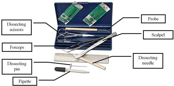
A dissection kit usually consists of the following instruments:
- knife or scalpel
- scissors (straight blades, curved blades)
- forceps, or tweezers (straight tip, curved tip)
- dissecting needles (straight, curved)
- dissecting pins
- dissecting probes (straight tip, curved tip)
- metric ruler
Directions: Watch Mussel Dissection to see how a dissecting kit is used to dissect a mussel.
During a dissection, scientists may use a dissecting microscope to view the specimen in greater detail. Dissecting microscopes are used to study objects in three-dimensions at low magnification. A dissecting scope has a stage where the specimen is placed. There may be a light source built in, or an external light source may be used. There is a single objective lens that can be zoomed in for a closer look by using the zoom adjustment knob. The specimen is viewed through binocular eyepieces.
Equipment for Biology: Mussel Cells
|
Like all animals, mussels develop from single cells (fertilized eggs). Mussels then develop into small larvae. A biologist who is studying mussel development will need to see the mussel larvae at all stages. Similarly, a biologist who is studying the anatomy or physiology of a mussel may want to see the cells that make up the mussel's tissues. To do so, the biologist will need a microscope. |

|
|
Compound Microscope 
|
This is a typical microscope that you may encounter in a biology classroom. It has a stage upon which a glass slide containing the specimen (cells, larvae, etc.) is placed. The mirror underneath is adjusted to shine light upward through the specimen. Some microscopes have a built-in light source or illuminator. The light is gathered by the objective lens on the nosepiece and transmitted through the tube to the eyepiece. The coarse and fine adjustment knobs help focus the image as you look through the eyepiece. This particular microscope is a monocular (one eyepiece) microscope, but there are also binocular (two eyepieces) microscopes as well. |
Equipment for Biology: Field Observations of Mussels
Investigations in biology often cannot be done in the classroom or in the laboratory. They must be done in the field. For example, mussels do not live alone but grow in communities or beds on the shoreline. To monitor populations, biologists measure the density of mussels on the shoreline (i.e., the number of mussels per unit area). One way to do this measurement is to use a quadrat. A quadrat is a square device of known area that is randomly placed throughout the muscle bed. The biologist then counts the number of mussels that lie within the quadrat. The photo below shows a biologist counting the mussel density on the shoreline with a quadrat.

Other devices that might be used in the field include a compass or global positioning satellite (GPS) receiver to note the precise location of mussel beds. Biologists might also use cameras to document their observations with photographs, hand lenses to magnify objects so that more details can be viewed, or binoculars to make observations from far away.
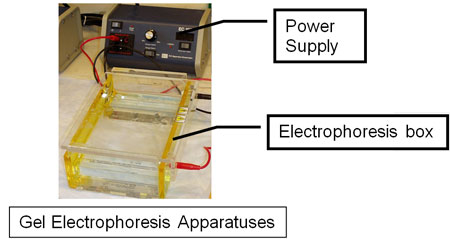
Equipment for Biology: Molecular Biology of Mussels—Gel Electrophoresis
In some studies of mussels, biologists may want to track genetic traits or specific mussel proteins. These types of studies involve molecular biology techniques like gel electrophoresis. Gel electrophoresis separates strands of DNA, RNA, or proteins from a mixed sample, based on size. It uses a gel, a chamber or box, and a power supply as shown below.
Directions: Explore how scientists use gel electrophoresis equipment by working through a virtual Gel Electrophoresis Lab. Follow the written steps by clicking the "Forward" arrow on the lower right of the graphic. Note how each piece of equipment is used.